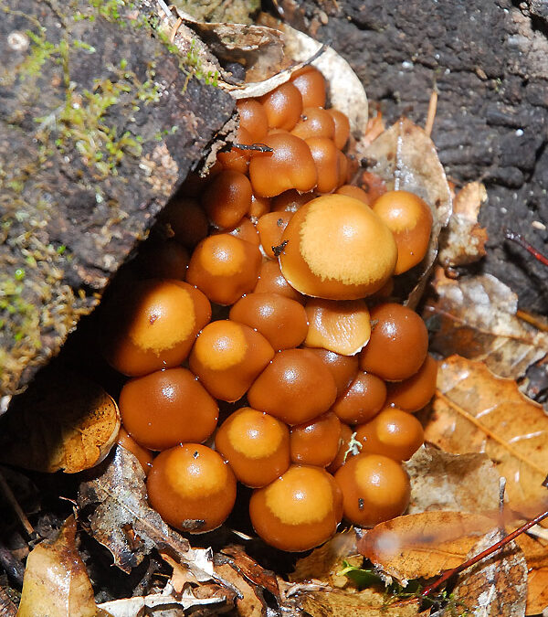
While we were installing the deer fence this past weekend, amidst some intermittent rain showers, we discovered a rather prolific colony of fungi surrounding an old decaying stump of a long since felled oak. At first we just noticed a few clusters of these tiny toadstools, but as we looked all around the stump, we were greeted with the most fascinating display of fungi we’ve seen here to date.

Although I am a biologist, I’m not a mycologist by any stretch of the imagination. My lack of mycological skills resulted in some significant head scratching and hair pulling when trying to identify this mysterious mushroom metropolis. My quest was simply to identify this monstrous mound of mushrooms that were encircling the base of the tree, purely out of curiosity. Little did I realize this was about to become a rather mammoth research effort. In the world of mushroom identification, one that I’ve now barely dunked my toes into, there seem to be some critical aspects to identifying the specimen in hand.

This species produces many impressive clusters of small fruiting bodies
First it is important to determine whether or not you’re looking at a gilled mushroom or not, which is easily achieved by flipping over a specimen and looking under the cap.

A look under the cap reveals this to be a gilled mushroom
Obviously cap shape is important but, as we discovered, this can be variable throughout the life-stage of the fungus. Cap color is important, but in some species, this can vary simply with atmospheric moisture. Accurately determining whether or not you’re looking at a mature specimen, or newly emerging fungi is important too, as even in this immature mushroom mound there is significant variability in both shape and color.

Some of the smallest of these mushrooms appear more spherical in shape, and darker brown

As they grow these mushrooms gradually reveal their stems, and a narrow white veil is visible at the cap margin

As the mushrooms mature, they take on a more classic sightly convex cap shape, and shielded from the wet weather, the cap color appears lighter and more opaque
The stem shape, and the presence or absence of any rings, color changes, and surface texture can also all help to provide clues as to the species. The challenge lies in the zillions of species of fungi out there, and separating our specimen from the herd!
After some delving into various texts and online resources I started to amass a list of potential candidates. At first I thought we may have found Gymnopus dryophilus. Although superficially this species seems similar to our mushroom mound, it was soon ruled out based primarily on gill color. The deadly Galerina marginata (syn. Galerina autumnalis) seemed like a plausible candidate for a while, but again, the gill color of our mushrooms was more a dusky brown than yellow. Bolbitius vitellinus had a similar cap shape, when young, but the margin seemed too striated compared with our specimen. We were close at one point with Hypholoma fasciculare, and Hypholoma capnoides, but the cap color seemed too dark with our specimens, and stem more slender. So the search continued on.

Similar appearing species to the mushroom growing around our oak stump. Clockwise from top left Gymnopus dryophilus, Galerina autumnalis, Hypholoma fasciculare, Hypholoma capnoides (photographs above Wikimedia Commons).
Refusing to accept my find as just another “LBM” (little brown mushroom…the mycologist’s equivalent of the birder’s little brown bird, or LBB), out of sheer desperation, I started running through the photo galleries at Hunting for Mushrooms, as many of the images of fungi there are from around the San Francisco Bay Area, and I wondered if our mushroom might be among them.
I whizzed through the first gallery of images, and on to the next, when suddenly I happened upon the images for Psathyrella hydrophila. I was ecstatic! After hours of running through mycological keys, skimming online images and databases, had I finally found what I was looking for? It seemed likely. So I went digging through the literature some more. Psathyrella hydrophila, also known as Psathyrella piluliformis (preferred), or Hypholoma hydrophila is apparently distinguished by a caespitose (clustered) fruiting habit, check, usually at the base of hardwood stumps, check, the tissue is hygrophanous (meaning translucent when wet, opaque when dry), check, has a brown cap with appendiculate veil fragments (fragments clinging to the cap margin) that in age become colored with maturing spores, check and check, and a white, fragile stem. Sounds promising!

These mushrooms were identified as Psathyrella piluiformis
My previous assumption that we might have a colony of deadly Galerina was further ruled out by reading in an online description of Psathyrella piluliformis that “Galerina autumnalis, a mushroom containing deadly amatoxins, is similar in color and size, but has a viscid/sticky cap when moist, the cap margin is typically striate, not appendiculate, and the spores are rusty-brown rather than dark brown.” Our mushroom, especially in the younger specimens, clearly is appendiculate. To bolster our confidence in identifying this mushroom as Psathyrella hydrophila/piluliformis, there was one last item worthy of checking for confirmation, spore color.
To determine the color of the spores, mycologists typically make spore prints. Samples of the mushroom in question are gathered, and the stems removed. The cap, with the gills or pores facing downward, is placed on a piece of paper. Not knowing if our mushroom produces white or colored spores, I placed samples on both white, and colored paper, fearing white spores might not show up on standard printer paper. I covered the samples on the paper with an upturned glass bowl, both to protect from drafts, and to protect my pets so they didn’t inadvertently consume my small science project, and then waited overnight to see what developed…

Spore Print from our Mystery Mushroom
As you can see, the spores are dark brown, just as they should be for Psathyrella piluliformis. Not too bad for my first spore print! I’m reasonably confident that we’ve identified this species correctly, but we’d welcome an expert opinion if there is one.
If there is anything important to be learned from this little foray into the fascinating world of fungus, it’s that absolute identification of many fungi is difficult at best, even for the experts. A minor error in judgement can result in catastrophic consequence. To the amateur eye, one mushroom may seem perfectly harmless, but in fact turn out to be deadly poisonous. As amateurs ourselves we of course know better than to consume any wild mushrooms we encounter. However, taking photographs and making spore prints is relatively harmless, and rather fun.
As the rainy season will be with us for the next few months, and our woodland areas have lots of damp dark areas full of decaying plant matter, I’ll have to take my camera out hunting again soon and see what other fascinating fungi I find flourishing here.
————————————————————–
Useful websites for identifying mushroom species:


I must be honest with you that I would have settled with the “little brown mushroom”. You have truly made your point concerning poisoned mushrooms. One can never be sure what is & what is not when it comes to edible ones.
Thanks for sharing this.
Isn’t nature amazing at the various methods it uses to decompose organic matter? In my Plant Biology class, I remember looking at many different types of fungi, but have forgotten what most of them are. I am amazed at the amount of different kinds of fungi present in the garden.
Wow! you are a detective!
Very educative post.Interesting! You did a great job! I used to be a mushroom hunter long ago. I had books, posters and the most important – a good teacher, my aunt. Many people loved to pick mushrooms and eat them. Delicious! I don’t do it now. Your pictures are great!Thank you!
Great post! Your reference to LBM, and LBB reminds me of DYC (damn yellow comps). Next time a mushrooms grows in my garden I am going to play detective too.